Talks
2026
Rapid advances in AI have expanded the range of capabilities required for successful real-world deployment. Understanding where we are in this multi-dimensional frontier is essential for accelerating innovation through effective quality assurance. Rigorous evaluation is increasingly difficult to scale as development requires testing many checkpoints across numerous benchmarks. Model comparison is further complicated by limited transparency of reported results. This talk explores challenges, best practices, and open-source tools that elevate evaluation to a core component of LLM development, delivering continuous signals across the model lifecycle.
We discuss principles for standardizing evaluation methods and improving consistency through practical patterns and anti-patterns, and examples of integrating the science of evaluation directly into model development. Using Nemo-Evaluator, an open-source scalable evaluation tool, we demonstrate modular architectures that enable transparent, reproducible measurement. Finally, we show how Nemo-Evaluator supports reproducible evaluation for the Nemotron model family, helping enable one of the most open development processes in modern AI.
2019
Mimicking radiologists to improve the robustness of deep-learning based automatic liver segmentation
RSNA 2019 Chicago, USA
[poster]
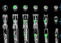
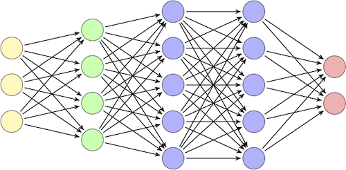
Radiologists delineating organ contours on a CT slice typically consider a couple of neighboring slices while taking into account the whole in-plane context in order to distinguish the organ boundary from surrounding structures. We present a new 3D deep-learning model that mimics the way radiologists interpret images on the example of liver segmentation. To evaluate its performance, the model is compared with a standard 3D neural network.
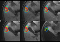
Explainability of decisions made by deep neural networks is of high value as it allows for validation and improvement of models. This work proposes an approach to explain semantic segmentation networks by means of layer-wise relevance propagation. As an exemplary application, we investigate which MRI sequences are most relevant for liver tumor segmentation.

KI in der Radiologie: Aktuelle Forschungsthemen
Invited talk
KIS-RIS-PACS und DICOM-Treffen, Schloß Waldthausen Mainz / Budenheim, Germany
[slides]
2018
Deep Learning Algorithms for Liver and Tumor Segmentation
Invited talk
SIRTOP Workshop @ Jahreskongress der TGRN e.V. und SRG e.V. in Kooperation mit HGMS e.V., Weimar, Germany
[slides]
Deep learning algorithms have the capability of automating many tedious and time-consuming medical image analysis tasks. In this talk a brief introduction into deep learning will be given as well as an overview of AI applications in the radioembolization context. Moreover, a study regarding automatic deep learning based liver and tumor segmentation in MRI and CT will be presented.
Automatic Liver and Tumor Segmentation in Late-Phase MRI Using Fully Convolutional Neural Networks
CURAC 2018, Leipzig, Germany
[slides]
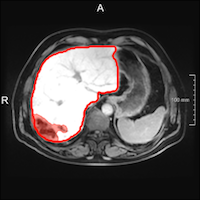
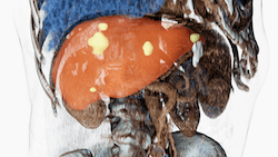
Liver and tumor segmentation plays an important role for many liver interventions. Automation of segmentation steps will bring a substantial improvement to clinical workflows by reducing the planning time and decreasing the inter-observer variability. While liver and tumor segmentation in CT data is a well-studied subject, only few publications are available for MRI data. We present an automatic segmentation approach based on 2D fully convolutional neural networks, which can be applied to both segmentation tasks not only in CT, but also in MR images. For the challenging MRI data, we evaluated two algorithms, one using an axial network and a second one based on three orthogonal models. The latter and better method resulted in a mean Dice index of 0.95 and 0.65 for liver and tumor segmentation, respectively. The liver segmentation quality of the method matches that of experienced clinical users and is higher compared to other automatic approaches and results in the literature. The tumor segmentation requires further improvements to make it comparable to the results of human experts.
Deep neural networks have significantly changed the way we design image processing pipelines. Thanks to their ability to accumulate knowledge from big data sets, neural networks are the core of many state-of-the-art computer vision algorithms. In this talk a brief history of computer vision will be given and examplary medical applications will be presented.
2017
Neural Network Based Automatic Liver and Liver Tumor Segmentation
LiTS Workshop @ MICCAI 2017, Quebec, Canada
[slides]
We present a fully automatic method employing fully convolutional neural networks (FCNNs) and a random forest (RF) classifier to solve the segmentation problems of the 2nd round of the Liver Tumor Segmentation Challenge (LiTS). In order to identify the ROI in which the tumors could be located, a liver segmentation is performed first. For the organ segmentation, an ensemble of FCNNs based on the U-net architecture is trained. Inside of the liver ROI, a 2D FCNN identifies tumor candidates, which are subsequently filtered with a random forest classifier yielding the final tumor segmentation. Our method ranked 3rd according to the segmentation evaluation.
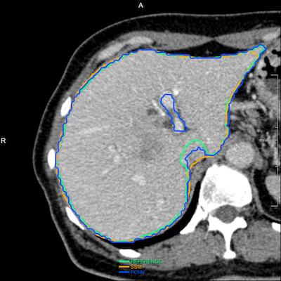
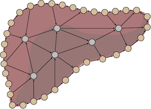
Many liver interventions require an organ segmentation for volumetry and procedure planning. The liver’s varying appearance in CT images makes this organ very time-consuming for manual delineation and challenging for automatic segmentation approaches. Automatic methods are desired, since they allow for a speed-up and reproducibility of the planning process. We investigated two automatic segmentation algorithms based on fully convolutional neural networks (FCN) and statistical shape models (SSM).

Comparison of Model Initialization Methods for Liver Segmentation using Statistical Shape Models
CARS 2017, Barcelona, Spain
[abstract]